Google

Mesothelioma !!

Also called: Malignant mesothelioma

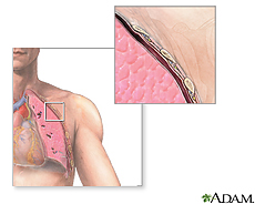
The tissue that lines your lungs, stomach, heart and other organs is called mesothelium. Mesothelioma is cancer of that tissue. It is a rare but serious type of cancer. It usually starts in the lungs, but can also start in the abdomen or other organs. Most people who develop mesothelioma have worked on jobs where they inhaled asbestos particles. It can take a long time - 30 to 50 years - between being around asbestos and getting the disease. Treatment includes surgery, radiation, chemotherapy or all three.

National Cancer Institute

Start Here
Basics Learn More Multimedia & Cool Tools
  • No links available
Research Reference Shelf For You
  • No links available

How Is Malignant Mesothelioma Staged?

Staging is the process of finding out how far the cancer has spread. Staging of mesothelioma is based on imaging studies such as x-rays, CT scans, and MRI scans. The treatment and outlook for patients with mesothelioma largely depends on the stage (extent of spread) of their cancer. Since pleural mesothelioma occurs most frequently and has been studied the most, it is the only mesothelioma for which a staging classification exists.

The major staging system has recently been developed by the International Mesothelioma Interest Group and adopted by the American Joint Committee on Cancer (AJCC). This is a TNM system, similar to staging systems used for most other cancers. T stands for tumor (its size and how far it has spread to nearby organs), N stands for spread to lymph nodes, and M is for metastasis (spread to distant organs). In TNM staging, information about the tumor, lymph nodes, and metastasis is combined in a process called stage grouping to assign a stage described by Roman numerals from I to IV.

T Stages

T1: Mesothelioma involves either the right or left pleura lining the chest. It has only spread to the pleura covering the lung, with the exception of possibly a few other small spots.

T2: Mesothelioma involves either the right or left pleura lining the chest and has spread from the lining of the chest into 1) the outer lining of the lung, 2) the diaphragm, or 3) into the lung itself.

T3: Mesothelioma involves either the right or left pleura lining the chest and has spread into 1) the first layer of the chest wall, 2) the fatty part of the mediastinum, 3) a single place in the chest wall, or 4) the outer covering layer of the heart.

T4: Mesothelioma involves either the right or left pleura lining the chest and has spread 1) into the chest wall, either muscle or ribs, 2) through the diaphragm, 3) into any organ contained in the mediastinum (esophagus, trachea, thymus, blood vessels), 4) into the spine, 5) across to the pleura on the other side of the chest, 6) through the heart lining or into the heart itself, or 7) into the brachial plexus (nerves leading to the arm).

N Stages

N0: No spread to lymph nodes.

N1: Spread to lymph nodes on the same side of the chest as the mesothelioma.

N2: Spread to lymph nodes around the point where the windpipe branches into the left and right bronchi or to lymph nodes in the space behind the chest bone and in front of the heart (mediastinum). Affected lymph nodes are on the same side of the cancerous lung.

N3: Spread to lymph nodes near the collarbone on either side, to hilar or mediastinal lymph nodes on the side opposite the cancerous lung.

M Stages

M0: No spread to distant organs or areas.

M1: The cancer has spread distantly.

Stage Grouping for Pleural Mesothelioma

Once the T, N, and M categories have been assigned, this information is combined (stage grouping) to assign an overall stage of I, II, III, or IV. Patients with lower stage numbers have a better prognosis.

Stage I (T1, N0, M0): Mesothelioma involves either the right or left pleura lining the chest. It has only spread to the outer lining of the lung in, at most, a few small spots. It has not spread to the lymph nodes or distant sites.

Stage II (T2, N0, M0): Mesothelioma involves either the right or left pleura lining the chest and has spread from the lining of the chest into 1) the outer lining of the lung, 2) the diaphragm, or 3) into the lung itself. It has not spread to the lymph nodes or distant sites.

Stage III (T1 or 2, N1 or 2, M0; OR T3, N0-2, M0): Mesothelioma involves either the right or left pleura lining the chest and may or may not have spread from the lining of the chest into 1) the outer lining of the lung, 2) the diaphragm, 3) into the lung itself and has spread to lymph nodes anywhere in the chest on the same side as the tumor, but has not spread to distant sites; OR Mesothelioma involves either the right or left pleura lining the chest and has spread into 1) the first layer of the chest wall, or 2) the fatty part of the mediastinum, or 3) a single place in the chest wall or 4) the outer covering layer of the heart and may or may not have spread to lymph nodes but not as far as to lymph nodes near the collarbone or on the opposite side of the chest. It has not spread to distant sites.

Stage IV (T4, any N, M0; OR any T, N3, M0; OR any T, any N, M1): Mesothelioma involves either the right or left pleura lining the chest and has spread 1) into the chest wall, either muscle or ribs, 2) through the diaphragm, 3) into any organ contained in the mediastinum (esophagus, trachea, thymus, blood vessels), 4) into the spine, 5) across to the pleura on the other side of the chest, 6) through the heart lining or into the heart itself, or 7) into the brachial plexus (nerves leading to the arm), and may or may not have spread to lymph nodes anywhere, but has not spread to distant sites; OR the tumor is of any size, but has spread to lymph nodes near the collarbone on either side, to hilar or mediastinal lymph nodes on the side opposite the cancerous lung but not to distant sites; OR the mesothelioma has spread to distant sites.

Other Prognostic Factors

Although stage is an important factor that determines a patient�s prognosis, other factors should also be considered. Some of these are poor performance status (for example being too sick to perform normal tasks of daily life), chest pain, shortness of breath, weight loss, high levels of a substance in the blood called LDH, low red blood cell count, high white blood cell count and others. These are considered serious factors and most people with all of these factors usually die within 6 months. Few live 2 years. Most people with none of these serious factors will live at least one year and have a 40% chance of living 2 years.

Cancer treatments

This page has links to the main radiotherapy and main chemotherapy sections, and to information about specific cancer drugs.

There is also a link to brief, general treatment pages from our About Cancer section. Below that, there is a list of links to the treatment sections for all specific cancers covered on CancerHelp UK. There is also information on treating symptoms and side effects is in its own section elsewhere on the site.


Chemotherapy
Radiotherapy
Hormone therapy
Biological therapy
Bisphosphonates
Individual cancer drugs
Ways of taking drugs

This link below will take you to a section of brief, general treatment information pages from our About Cancer section.
About cancer treatments

Treating specific cancers.


There are links below to the treatment sections for all the specific cancers covered on CancerHelp UK.

Treating acute leukaemia
Treating bladder cancer
Treating bone cancer
Treating bowel cancer
(colorectal cancer)

Treating brain tumours
Treating breast cancer
Treating cervical cancer
Treating chronic leukaemia
Treating gallbladder cancer
Treating Hodgkin's disease
Treating kidney cancer
Treating larynx cancer
Treating liver cancer
Treating lung cancer
Treating melanoma
Treating mesothelioma
Treating mouth and oropharyngeal cancer
Treating multiple myeloma
Treating nasal and sinus cancers
Treating nasopharyngeal cancer
Treating non-Hodgkin's lymphoma
Treating oesophageal cancer
Treating ovarian cancer
Treating pancreatic cancer
Treating prostate cancer
Treating skin cancer
(not melanoma)

Treating soft tissue sarcoma
Treating stomach cancer
Treating testicular cancer
Treating thyroid cancer
Treating vaginal cancer
Treating vulval cancer
Treating unknown primary cancer
Treating womb cancer

Information about other cancer treatments will be added over time


*more infoForward to Chemotherapy


Last updated 29 January 2007

CancerHelp UK is not designed to provide medical advice or professional services and is intended to be for educational use only. The information provided through CancerHelp UK is not a substitute for professional care and should not be used for diagnosing or treating a health problem or a disease. If you have, or suspect you may have, a health problem you should consult your doctor.

Diagnosing mesothelioma

This section tells you about the tests used to diagnose mesothelioma. You can choose from the following menu

Tests for mesothelioma
Further tests for mesothelioma
Questions for your doctor about tests for mesothelioma


Other information you may find useful

Reading list
Help and support
On-line services


Back to Questions for your doctor about mesotheliomamore infoForward to Tests for mesothelioma


Last updated 13 April 2007

CancerHelp UK is not designed to provide medical advice or professional services and is intended to be for educational use only. The information provided through CancerHelp UK is not a substitute for professional care and should not be used for diagnosing or treating a health problem or a disease. If you have, or suspect you may have, a health problem you should consult your doctor.

犬惡性間皮細胞瘤

一、 病歷:
  患犬為一隻體重21.7 kg之混種閹公犬,主訴為外觀消瘦,腹部膨大,呼吸困難及精神沈鬱等症狀,由地方動物醫院轉診至本校教學醫院就診。
  臨床檢查:經放射線及超音波均發現胸腹腔皆有大量液體蓄積,且於肝、脾臟表面可見圓形或卵圓形疑似腫瘤團塊。投予抗腫瘤藥物無明顯改善。
  胸、腹水檢查在超音波影像引導下實施胸、腹腔穿刺術,抽取 胸、腹腔內液體進行檢查。胸、腹腔內液體皆為深紅色、血樣、混濁、無臭、不具黏稠性及凝固性,總有核細胞數2.1×10³/μl,蛋白質含量0.5 g/dL,比重1.015,為修飾的漏出液(modified transudate)。抹片鏡檢下以紅血球為主,還有少數反應型間皮細胞、吞噬細胞及非退行性白血球。
  多次抽取血樣胸腹水約800~3500 ml。且進行兩次開腹探測,並於橫膈、腹壁、胃、肝、脾臟及胃網膜可見有多發,直徑0.5-3.0公分,大小不一,表面光滑的圓形或卵圓形疑似腫瘤的團塊。
  進行生檢,診斷為間皮細胞瘤。最後於治療期間死亡,移送病理診斷。
二、
肉眼病變:
  該犬外觀消瘦脫水,脂肪呈嚴重漿液性萎縮,背部皮下脂肪消失。胸腔與腹腔中皆有大量 之血樣液體。並可見胸壁,心包囊漿膜面,心外膜及橫膈表面瀰漫性的佈滿白色表面粗糙之小結節。在腹壁、胃、腸系膜、腸系膜淋巴結、肝臟、脾臟、膀胱與左腎 被膜表面,皆可見白色大小不一表面光滑觸感堅實之團塊(圖1,2,3)。其他病變如下:
1.
心臟:右心房中可見兩條心絲蟲成蟲,左心可見心內膜增殖,左右心腔皆可見血液凝塊。
2.
肺臟:肺臟有壓迫萎縮現象,右肺膈葉與胸壁黏連。
3.
胃大彎、大網膜:成團塊樣。
4.
膀胱:內有粉末結晶。
5.
脾臟:萎縮。
三、 組織病變:
1.
肝臟:有少量血鐵素沉著。肝臟腫瘤團塊,低倍下內為乳突狀似腺管樣結構內含粉紅色均質物,外有結締組織包被。腫瘤細胞之細胞核大,形狀為圓到卵圓形,核質比大於一,有絲分裂相當明顯,疑似為上皮性之腫瘤。
2.
肺臟:輕微水腫與鬱血,肋膜之結締組織增厚,且肺臟與肋膜交界處有腫瘤團塊,其型態同上。
3.
橫膈:肌纖維有腫瘤細胞浸潤。
4.
腎臟:間質性腎炎,皮質部炎症細胞浸潤,以單核細胞為主。絲球體內有粉紅色物質
沈著;且可見相同型態之腫瘤團塊。
5.
腸系膜:腫瘤團塊內為乳突狀似腺管樣結構內含粉紅色均質物,外有結締組織包被。腫瘤細胞之細胞核大,形狀為圓到卵圓形,有絲分裂相當明顯(圖4)。
四、 診斷:
犬惡性間皮細胞瘤(Malignant Mesothelioma in a Dog)
五、 討論:
  間皮細胞瘤在狗貓之發生率約 為千分之一,且十分少見。腫瘤細胞主要侵犯胸腹腔與心包囊,也有侵犯至陰囊之vaginal tunic。依據人的流行病學調查結果,暴露於含石綿之環境中,為致病的重要因素之一,其他如貴橄欖石(chrysolite)、角閃石 (Amphibole)等其他礦物質纖維皆為致癌因子,皆能有效地誘導染色體1、2、3、6、11、17及22重組,導致proto-oncogenes 的活化與tumor suppressor genes的不活化,但詳盡的致病機制仍不清楚。依據Glickman等人於1983年發表的報告中指出,發病的患犬,其畜主可能常工作於石綿環境中,或 是與殺蟲劑的使用,驅蚤劑的污染有關。當巨噬細胞吞噬石綿纖維後,產生之oxygen-free radical會重複的造成tumor precursor cell的基因性傷害,導致化生。經石綿纖維活化之巨噬細胞同時分泌IL-1、FGF(fibroblast growth factor)、PDGF使細胞增殖。
  組織學上,正常之間皮層為一單 層細胞之間皮細胞。間皮細胞表面含microvilli、desmosome並具有吞噬與分泌的能力。體腔內的炎症與傷害,可使間皮細胞產生明顯的生理性 增殖;體液的蓄積也會造成間皮細胞的脫落與生成。間皮細胞型態為扁平或立方上皮細胞,但由中胚層分化而來。因此間皮細胞瘤可呈現上皮性、纖維性或混合性之 組織型態。間皮細胞瘤因可於體腔內散播,並造成多發之腫瘤生長而視為惡性瘤。
  典型的間皮細胞瘤為瀰漫性分布於體腔壁上或臟器漿膜面之結節團塊,由於腫瘤表面的分泌物與因腫瘤組織阻塞之淋巴管而造成廣泛性之體液蓄積,常見之臨床症狀為呼吸困難、胸腹水與腹部膨大。若有侵犯至心包囊之間皮細胞瘤則可能伴隨著急性血栓與右心衰竭。
  在組織型態上,上皮性的主要特 徵為組織長成乳頭狀及管狀的型態,通常主要是立方上皮細胞,其次為扁平型,只有很少數的細胞型態為柱狀上皮細胞;細胞質有單一至多個圓型的細胞質空泡,細 胞核大,核仁也大;細胞表面可見有刷狀緣(adenocarcinoma則是以柱狀為主,且無brush border)。纖維性間皮細胞瘤的細胞型態為卵圓形、長形或紡錘形,同時也如上皮性一般具有vesicular nuclei。
  在診斷方面,胸腹水之細胞學檢 查無法有效地診斷出間皮細胞瘤,雖然在胸腹水中可見到脫落之間皮細胞,但生理上的增殖與化生卻是難以區分的;其他的檢查如放射線學、超音波、CT或是 MRI皆只是輔助診斷,而直接生檢是最直接診斷的方式。典型的病例以H&E染色配合臨床表現即可確診,否則需要免疫化學染色來幫助確診。
六、 參考文獻:
1.
Barrett JC. Chromosomal mutation and asbestos carcinogenicity. In: Cellular and molecular
aspects of fiber carcinogenesis. Harris CC, Lenchner JF, and Brinkley BR. pp 36-37, 1991.
2.
Caminschi I, Venetsanakos E, Leong CC, Garlepp MJ, Robinson BWS, and Scott B. Cytokine
gene therapy of mesothelioma. Immune and antitumor effects of transfected interleukin-12.
Am J Respir Cell Mol Biol 21:347-356, 1999.
3.
Closa. JM, Font A, and Mascort J. Pericardial mesothelioma in a dog :long-term survival after
pericardiectomy in combination with chemotherapy. J S A P 40:383-386, 1999.
4.
Glickman LT, Domanski LM, Maguire TG, Dubielzig RR, and Churg A. Mesothelioma in pet
dogs associated with exposure of their owners to asbestos. Environ Res 32:305-313, 1983.
5.
Jaurand MC, Bignon J. The mesothelial cell and mesothelioma. Lung Biology in Health and
Disease. 787 : 71-297, 1994.
6.
Jones TC. Hunt RD, and King NW. In:Veterinary pathology. pp 972-973, 992-993, 1997.
7.
Jubb KVF, Kennedy PC, and Palmer N. Neoplastic diseases of the peritoneum. In:Pathology
of domestic animals. 4th ed. Vol.2. pp 443-444, 1993.
8.
Roberts JR. Surgical treatment of mesothelioma: pleurectomy. Chest. 116:446s-449s, 1999.
9.
Smith DA, and Hill FWG. Metastatic malignant mesothelioma in a dog. J Comp Pathol 100(1)
:97-101, 1989.
圖1:胸腔與腹腔中皆有大量之血樣液體,並佈滿白色表面粗糙之圓球形小結節。

圖2:在腹壁、胃、腸系膜、腸系膜淋巴結,佈滿白色表面粗糙之圓球形小結節。直徑0.1 cm至1.0 cm均有。

圖3:肝臟、脾臟、膀胱與左腎被膜表面,皆可見白色大小不一表面光滑觸感堅實之團塊。

Mesothelioma


Original Article:http://www.nlm.nih.gov/medlineplus/mesothelioma.html
Mesothelioma
Introduction

Malignant mesothelioma is a rare cancer that occurs in the thin layer of tissue that covers the majority of your internal organs (mesothelium). Doctors divide mesothelioma into different categories based on what part of the mesothelium is affected by cancer. Mesothelioma that occurs in the tissue that surrounds the lung (pleura) is called pleural mesothelioma and is the most common form. Mesothelioma that occurs in the tissue in your abdomen (peritoneum) is called peritoneal mesothelioma and accounts for 10 percent to 20 percent of all mesotheliomas. In rare cases, mesothelioma can also occur in the lining around the heart (pericardium) and in the lining around the testicles (tunica vaginalis).

Between 2,000 and 3,000 people are diagnosed with mesothelioma in the United States each year. Men are more likely to develop mesothelioma than are women. It's more common in older adults — most people with mesothelioma are 65 and older — though mesothelioma can be diagnosed at any age.

Mesothelioma is closely linked to exposure to asbestos — a natural fiber that was once used in manufacturing a wide variety of industrial and household products. Mesothelioma rates have increased during the past 20 years in response to the widespread use of asbestos in the past. Many industrialized countries now limit asbestos use and enforce laws to protect workers who may be exposed. Researchers predict these efforts will lead to fewer cases of mesothelioma in the future.
Signs and symptoms

Signs and symptoms of mesothelioma vary depending on where the cancer occurs.

Pleural mesothelioma signs and symptoms may include:

* Shortness of breath
* Painful breathing
* Chest pain under the rib cage
* Unusual lumps of tissue under the skin on your chest
* Unexplained weight loss
* Dry (nonproductive) cough

Peritoneal mesothelioma signs and symptoms may include:

* Abdominal pain
* Abdominal swelling
* A change in your bowel habits, such as more frequent diarrhea or constipation
* Lumps of tissue in the abdomen
* Unexplained weight loss

Signs and symptoms of pericardial mesothelioma and mesothelioma of the tunica vaginalis are unclear. These forms are so rare that not much information is available. Mesothelioma of the tunica vaginalis may be first detected as a mass on a testicle. Pericardial mesothelioma signs and symptoms may include difficulty breathing and fever.

Signs and symptoms of mesothelioma that has spread to other parts of the body include:

* Pain in the area where cancer has spread
* Difficulty swallowing
* Swelling in the neck and face

Causes

In general, cancer begins with a genetic mutation that turns normal, healthy cells into abnormal cells. Healthy cells grow and multiply at a set rate, eventually dying at a set time. Abnormal cells grow and multiply out of control, and they don't die. The accumulating abnormal cells form a mass (tumor). Cancer cells invade nearby tissues and can break off from an initial tumor to spread elsewhere in the body (metastasize).

It isn't clear what causes the initial genetic mutation that leads to mesothelioma, though researchers have identified factors that may increase the risk. It's likely that cancers form because of an interaction between many factors, such as inherited conditions, your environment, your health conditions and your lifestyle choices.
Risk factors

Asbestos exposure plays a role in 70 percent to 80 percent of mesothelioma cases, though the actual percentage could be higher. Asbestos is a mineral that is found naturally in the environment. Asbestos fibers are strong and resistant to heat, making them useful in a wide variety of applications. Asbestos fibers have been used to make insulation, cement, brakes, shingles, flooring and many other products.

People who work around asbestos fibers are thought to have the greatest risk of mesothelioma. When asbestos is broken up — for instance, in the mining process or in removing asbestos insulation from a building — dust may be created. If the dust is inhaled or swallowed, the asbestos fibers may settle in the lungs or in the stomach, where they can cause irritation that may lead to mesothelioma, though how exactly this happens isn't understood. Although asbestos is still used in a limited number of industries, the federal government limits the amount of asbestos fibers workers may be exposed to and sets rules to protect workers.

Mesothelioma risk is believed to be increased in people who are exposed to high levels of asbestos, in people who are exposed to asbestos over a long period of time and in people exposed to asbestos at a young age. It can take 30 to 40 years or more for mesothelioma to develop as a result of asbestos exposure.

People who live with workers exposed to asbestos may also have an increased risk of mesothelioma. Asbestos dust is thought to have been carried home on workers' clothes. Today workers are required to shower and change clothes after working with asbestos to protect their families.

Some people with years of asbestos exposure never develop mesothelioma. Researchers estimate only about 5 percent of the people with the highest exposure to asbestos will develop mesothelioma. And yet, others with very brief exposure develop the disease. This indicates that other factors may be involved in determining whether someone gets mesothelioma or doesn't. For instance, you could inherit a predisposition to cancer or some other condition could increase your risk. Risk of mesothelioma is increased greatly in smokers who are exposed to asbestos.

Other possible risk factors
Researchers have identified other factors that may increase the risk of mesothelioma, including:

* SV40. Some research indicates a link between mesothelioma and simian virus 40 (SV40), a virus originally found in monkeys. Millions of people may have been exposed to SV40 when receiving polio vaccinations between 1955 and 1963, because the vaccine was developed using monkey cells. Once it was discovered that SV40 was linked to certain cancers, the virus was removed from the polio vaccine. There is some evidence that SV40 may also be passed between people, though it isn't clear how. Whether SV40 increases the risk of mesothelioma is a point of contention, and more research is needed.
* Radiation. The radioactive substance thorium dioxide was used along with X-rays to diagnose various health conditions from the 1920s to the 1950s. Thorium dioxide was later found to cause cancer and was no longer used. Some research links thorium dioxide to mesothelioma.
* Asbestos-like minerals. A naturally occurring asbestos-like mineral called zeolite has been linked to mesothelioma cases in Turkey, where the mineral is used to construct homes.
* Family history. Research into the same region of Turkey where zeolite is used reveals that family history may play a role in mesothelioma there. More research is needed to determine whether family history may predispose some people to mesothelioma.

When to seek medical advice

See your doctor if you have signs and symptoms that may indicate mesothelioma. Signs and symptoms of mesothelioma aren't specific to this disease and may be related to other conditions. If any signs and symptoms seem unusual or bothersome to you, ask your doctor to check them out.
Screening and diagnosis

If you have signs and symptoms that might indicate mesothelioma, your doctor will conduct a physical exam, paying particular attention to areas where you're experiencing pain. He or she checks for any lumps or other unusual signs. Your doctor may order other tests to determine the cause of your signs and symptoms, including:

* Chest X-ray. X-rays may show abnormalities if you have pleural mesothelioma.
* Chest or abdominal CT scan. Computerized tomography (CT) may reveal abnormalities in your chest or abdomen if you have mesothelioma.

It's not uncommon for mesothelioma to be misdiagnosed initially because mesothelioma is rare and its signs and symptoms aren't specific. Your doctor will likely rule out other more common conditions before considering mesothelioma.

Biopsy
Biopsy, a surgical procedure to remove a small portion of the mesothelium for laboratory examination, is the only way to determine whether you have mesothelioma. Depending on what area of your body is affected, your doctor selects the right biopsy procedure for you. Options include:

* Fine-needle aspiration. The doctor removes fluid or a piece of tissue with a small needle inserted into your chest or abdomen.
* Thoracoscopy. Thoracoscopy allows the surgeon to see inside your chest. In this procedure, the surgeon makes one or more small incisions between your ribs. He or she inserts a tube with a tiny video camera to see inside your chest cavity — a procedure sometimes called video-assisted thoracoscopic surgery (VATS). Special surgical tools allow your surgeon to cut away a piece of tissue.
* Laparoscopy. Laparoscopy allows the surgeon to see inside your abdomen. Using one or more incisions into your abdomen, the surgeon inserts a tiny camera and special surgical tools to obtain a small piece of tissue for examination.
* Thoracotomy. Thoracotomy is surgery to open your chest to allow a surgeon to check for signs of disease. He or she removes a sample of tissue for testing.
* Laparotomy. Laparotomy is surgery to open your abdomen to allow a surgeon to check for signs of disease. He or she removes a sample of tissue for testing.

Once the tissue sample has been collected through biopsy, the sample is analyzed under a microscope. This determines whether or not the abnormal tissue is mesothelioma. Biopsy samples also allow your doctor to test for the type of cells involved in your mesothelioma. The type of mesothelioma you have is used to determine your treatment plan.

Staging
Once mesothelioma is diagnosed, your doctor orders other tests to determine the extent of the cancer and whether it has spread — a process called staging. Imaging procedures allow doctors to see inside your chest or abdomen to determine the stage of mesothelioma. Options include:

* Chest X-ray
* CT scans of the chest and abdomen
* Magnetic resonance imaging (MRI)
* Positron emission tomography (PET)

Once the extent of mesothelioma is determined, a stage is assigned. Staging helps your doctor determine your prognosis and the best treatment plan. The stages of mesothelioma are:

* I. Stage I mesothelioma is considered localized cancer, meaning it's limited to one small area of the chest or abdomen.
* II. Stage II mesothelioma is considered advanced cancer. Mesothelioma at this stage involves the mesothelium and has also spread to other structures directly adjacent to the tumor, such as the lungs or the diaphragm.
* III. Stage III mesothelioma is also considered advanced cancer. Mesothelioma at this stage meets the same requirements as stage II, but has also spread to the lymph nodes in the region.
* IV. Stage IV mesothelioma is an advanced cancer that has spread to distant areas (metastasized). Mesothelioma most commonly spreads (metastasizes) to the brain and areas of the lung that are away from the tumor.

Complications

As pleural mesothelioma spreads in the chest, it puts pressure on the structures in that area. This can cause complications, such as:

* Difficulty breathing
* Chest pain
* Difficulty swallowing
* Swelling caused by pressure on the large vein that leads from your upper body to your heart (superior vena cava syndrome)
* Pain caused by pressure on the nerves and spinal cord

Mesothelioma that progresses can lead to death. People who die of mesothelioma usually die from related complications, such as lung failure, bowel obstruction, heart problems, stroke and other causes.
Treatment

What treatment you undergo for mesothelioma depends on your health and certain aspects of your cancer, such as its stage and location. Unfortunately, mesothelioma often is an aggressive disease and for many people a cure won't be possible. Mesothelioma is usually diagnosed at an advanced stage — when it isn't possible to remove the cancer through surgery. Instead, your doctor may work to control your cancer and its signs and symptoms to make you more comfortable.

Discuss your treatment goals with your doctor. Some people want to do everything they can to treat their cancer, even if that means enduring side effects for a small chance of a cure. Others prefer treatments that make them comfortable so that they can live their remaining months as symptom-free as possible.

Mesothelioma treatment options may include surgery, radiation, chemotherapy and clinical trials.

Surgery
Surgeons work to remove mesothelioma from your body. Sometimes it isn't possible to remove all of the cancer. In those cases, surgery may help to reduce the signs and symptoms caused by mesothelioma spreading in your body. Surgical options may include:

* Surgery to decrease fluid buildup. Pleural mesothelioma may cause fluid to build up in your chest, causing difficulty breathing. Surgeons insert a tube or catheter into your chest to drain the fluid. Surgeons may also inject medicine into your chest to prevent fluid from returning (pleurodesis).
* Surgery to remove the tissue around the lung or abdomen. Surgeons may use scalpels and other surgical tools to remove the tissue lining the ribs and the lungs (pleurectomy) or the tissue lining the abdominal cavity (peritonectomy) in order to relieve signs and symptoms of mesothelioma.
* Surgery to remove as much of the cancer as possible (debulking). If all of the cancer can't be removed, surgeons may attempt to remove as much as possible.
* Surgery to remove a lung and the surrounding tissue. Removing the affected lung and the tissue that surrounds it may relieve signs and symptoms of pleural mesothelioma. This procedure also allows doctors to use higher doses of radiation against any remaining mesothelioma, since doctors won't need to worry about protecting your lung from damaging radiation.

Chemotherapy
Chemotherapy uses chemicals to kill cancer cells. Chemotherapy drugs travel throughout your body and kill rapidly growing cells. This works against cancer cells, but also affects other rapidly growing cells in your body, such as those in your hair follicles and those in your gastrointestinal system. Chemotherapy may slow the growth of pleural mesothelioma. Chemotherapy can be used before surgery (neoadjuvant chemotherapy) or after surgery (adjuvant chemotherapy) to reduce the signs and symptoms you may experience from mesothelioma.

People with peritoneal mesothelioma may receive adjuvant chemotherapy drugs that have been heated (hyperthermic chemotherapy). Rather than being distributed throughout the body, chemotherapy drugs are often injected directly into the abdominal cavity (intraperitoneal chemotherapy), where they can reach the peritoneal mesothelioma directly without injuring healthy cells in other parts of the body. This allows doctors to administer higher doses of chemotherapy drugs. Intraperitoneal chemotherapy may also be used to reduce the signs and symptoms of peritoneal mesothelioma that can't be removed through surgery.

Radiation therapy
Radiation therapy focuses high-energy radiation to a specific spot or spots on your body. Radiation may reduce signs and symptoms in people with pleural mesothelioma. Doctors aim radiation at the entire chest to obtain the best result. However, many sensitive organs reside in the chest, such as the heart, lungs and spinal cord, so doctors must use low doses of radiation to spare these organs. Radiation therapy is sometimes used after biopsy or surgery to prevent mesothelioma from spreading to the surgical incision.

Radiation therapy is used occasionally in people with peritoneal mesothelioma. Radiation may reduce signs and symptoms of mesothelioma.

Combination therapy
Surgery, chemotherapy and radiation therapy may be combined. This aggressive therapy can be grueling and may not be appropriate for everyone. Younger, healthier people and those with earlier stage mesothelioma may be more able to endure this treatment. Combination therapy has shown the most promise in treating mesothelioma. However, most people will eventually experience a recurrence of this cancer despite this aggressive treatment. Combination therapy has been used in both pleural mesothelioma and peritoneal mesothelioma.

Clinical trials
Clinical trials are studies of new mesothelioma treatment methods. People with mesothelioma may opt for a clinical trial for a chance to try new types of treatment. However, a cure isn't guaranteed. Carefully consider your treatment options and talk to your doctor about what clinical trials are open to you. Your participation in a clinical trial may help doctors better understand how to treat mesothelioma in the future.

Treatment for other types of mesothelioma
Pericardial mesothelioma and mesothelioma of the tunica vaginalis are very rare and can be very aggressive. Early-stage cancer may be removed through surgery. Doctors have yet to determine the best way to treat later stage cancers, though. Your doctor may recommend other treatments to improve your quality of life.
Prevention

Reducing your exposure to asbestos may reduce your risk of mesothelioma. Most people with mesothelioma were exposed to the asbestos fibers at work. Workers who may encounter asbestos fibers include:

* Miners
* Factory workers
* Insulation manufacturers
* Railroad workers
* Ship builders
* Gas mask manufacturers
* Construction workers

Follow all safety precautions in your workplace, such as wearing protective equipment. You may also be required to shower and change out of your work clothes before taking a lunch break or going home. Talk to your doctor about other precautions you can take to protect yourself from asbestos exposure.

Older homes and buildings may contain asbestos. In many cases, it's more dangerous to remove the asbestos than it is to leave it intact. Breaking up asbestos may cause fibers to become airborne, where they can be taken into your body as you breathe. Consult experts trained to detect asbestos in your home. These experts may test the air in your home to determine whether the asbestos is a risk to your health. Don't attempt to remove asbestos from your home — hire a qualified expert.
Coping skills

Shock. Anger. Sadness. Despair. Confusion. You may be feeling one or all of these emotions after receiving a mesothelioma diagnosis. A cancer diagnosis is devastating not only to you, but to your family and loved ones. Take time to experience the sadness and despair and to grieve. As you're shuffled between appointments with various doctors and specialists, know that you can take control of the time you have remaining. You decide how you'll spend your time and whom you'll spend your time with. Along with your doctor, you determine which treatments you'll undergo.

In order to regain a sense of control, try to:

* Learn everything you can about mesothelioma. Write down a list of questions to ask your doctor. Ask your health care team for reading materials and other resources to help you better understand your disease. Consult the National Cancer Institute (NCI) and the American Cancer Society (ACS) for more information. Both organizations can provide you with information through their Web sites or toll-free telephone information lines. Call the NCI at 800-4-CANCER, or 800-422-6237. Call the ACS at 800-ACS-2345, or 800-227-2345. Information may help you feel more confident in your treatment decisions, and it may help you better understand what's going on inside your body.
* Surround yourself with a support network. Whether it's close friends or family or a combination of both, surround yourself with people who love you. These people can help you with the everyday tasks, such as getting you to appointments or treatment, and they can help support you by providing someone to talk to or to reassure you. If you have trouble asking for help, learn to be honest with yourself and accept help when you need it — your friends and family feel helpless too, and they want to help you.
* Seek out other people with cancer. Ask your health care team about cancer support groups in your community. Sometimes there are questions that can only be answered by other people with cancer. Support groups offer a chance to ask these questions and receive support from people who understand your situation. Online support message boards can offer similar benefits while allowing you to remain anonymous.
* Plan for the unknown. Ask your health care team about advance directives that give your family guidance on your medical wishes in case you can no longer speak for yourself. Talk to a lawyer about your will, if you haven't already done so.


By Mayo Clinic Staff
Aug 11, 2006
© 1998-2007 Mayo Foundation for Medical Education and Research (MFMER). All rights reserved. A single copy of these materials may be reprinted for noncommercial personal use only. "Mayo," "Mayo Clinic," "MayoClinic.com," "EmbodyHealth," "Reliable tools for healthier lives," "Enhance your life," and the triple-shield Mayo Clinic logo are trademarks of Mayo Foundation for Medical Education and Research.

DS00779
Mayo Clinic

Treatment

Treatment

* Chemotherapy for Malignant Mesothelioma(American Cancer Society)
* Pleurodesis(International Mesothelioma Program)
* Radiation Therapy for Malignant Mesothelioma(American Cancer Society)
* Surgery for Malignant Mesothelioma(American Cancer Society)

腹膜間皮癌(Peritoneal Mesothelioma)

1. 處理原則: 減量手術(debulking) → 化療 → 全腹腔電療


2. 化療處方: CA (CYC, ADR): 400, 40 mg/m2 D1, 8 q4w

整合治療(Combined-modality therapy) < 仍在調查中>

1. Sugarbaker DJ(J Thorac cardiovasc Surg 1991):
做法:EPP→ 4~6週後 → 輔助性化療 CAP(600,60,75/m2) q3w → ± 電療 55Gy
結果:31位病人,中位存活期16個月,3年存活率36%。有兩種情況的存活率增加:完全切除和手術邊緣乾淨。在epithelial cell-type的病人, 3年整體存活率42%; 但mixed or fibrosarcomatous cell or mediastinal LN+結果仍差。

但Rusch報告:EPP手術的存活期500天; 非EPP的手術存活期325天。所以EPP不能治癒; 而非EPP的手術也不會較差。


2. MSKCC: pleurectomy/decortication ± IORT(125I 或 192Ir) → 外電 45Gy
整體存活率和局部控制都很差; 中位存活期 24個月。


3. Weder W(JCO 2004):
做法:根據Byrne(JCO 1999)的報告,給予三個療程前導性化療(每28天為一週期,接受三個療程的gemcitabine 1000mg/m2 Day 1,8,15 + cisplatin 80mg/m2 Day 1→ EPP→ 輔助性電療
結果:19位病人,前導性化療反應率32%,中位存活期23個月。這個結果遠優於Taverna C(Ann Oncol 2000)的報告(EPP→輔助性化療→輔助性電療),其中位存活期只有13個月;也優於Sugarbaker DJ(J Thorac cardiovasc Surg 1999)的報告(EPP→輔助性化療→輔助性電療),其 中位存活期19個月。

腔內療法 (Intracavity therapy, IP)

1. 效果不佳, 因為藥物的穿透力差。


2. 主要的研究針對在輔助性治療(adjuvant)的情況(即手術 → 腔內化療 → 全身化療)。
a) Rusch: Pleurectomy後立即 IP CDDP(100mg/m2) + MMC(8mg/m2)

b) IFN-γ: 四千萬單位(40MU)輸注到肋膜腔, 一週兩次, 連續8週。在第一期且腫瘤小於5mm的病人, 反應率45%(4/9 CR)。

化學治療

單一藥物反應率只有13%, 沒有一個藥物有超過20%的反應率。複方化療反應率也只有14%, 並不會優於單一藥物。也就是說沒有有效的藥物(none active)。最具活性的種類是anthracyclines、platinums、antifolates。還有許多針對間皮癌的新藥最近也已經發展出來, 包括antifolates(pemetrexed和raltitrexed)、ribonuclease(ranpirnase)、vinorelbine、和gemcitabine。間皮癌獨特的生物學使得進行中對抗血管內皮生長因子(VEGF)、上皮生長因子(EGF)、和血小板衍生生長因子(PDGF)等的標靶藥物(targeted agents)的臨床研究特別令人感到興趣。


1. Anthracyclines:
a) Doxorubicin:
在歷史上被認為是惡性間皮癌的黃金標準治療。
根據ECOG 1983年的報告, Doxorubicin單一藥物的反應率只有14%。
Breau(1991): ADR + BLM + CDDP + MMC 總反應率 44%。
Sampson(1987): ADR + CYC ± DTIC 總反應率 12%。
CALGB(1993): CDDP + MMC和CDDP + ADR 的總反應率相似, 約14-26%。

b) Epirubicin:
Epirubicin的活性也和Doxorubicin差不多, Magri(Tumori 1991)每三星期給予75mg/m2的反應率5%, Mattson(JCO 1992)每三星期給予110mg/m2的反應率15%。

c) Liposomal doxorubicin: 反應率0~7%

d) Liposomal daunorubicin: 反應率0%


2. Platinum analogs:
a) Cisplatin:
SWOG(Invest New Drugs 1988)每三星期給予100mg/m2的整體反應率14%。 Planting(Ann Oncol 1994)每星期給予80mg/m2的部份反應率36%。

b) Carboplatin:
CALGB(1990)每四星期給予400mg/m2的反應率7%。

c) Oxaliplatin:
在間皮癌沒有單一藥物的研究。Fizazi(JCO 2000)的第一期研究將Oxaliplatin和raltitrexed併用於cisplatin無效的病人有某些活性。這結果導致Fizazi(Proc ASCO 2000)將這種Oxaliplatin和raltitrexed組合用於72位間皮癌, 其中有16位曾接受過化療, 結果反應率有26%, 如果是cisplatin無效的病人還有20%的反應率。

d) ZD0473:
Giaccone(Eur J Cancer 2001)對曾經治療過的病人沒有任何活性。


3. Antifolates(pemetrexed和raltitrexed):
a) Antifolates是對抗惡性間皮癌化療藥物中最具活性的種類。如下表:
參考文獻 藥物 病人數目 反應率(%)
Kindler(Cancer 1999) Edatrexate 20 25
Kindler(Cancer 1999) Edatrexate/Leucovorin 40 16
Solheim(Br J Cacner 1992) High-dose methotrexate 60 37
Scagliotti(Eur J Cancer 2001) Pemetrexed(Alimta®) 62 14.5
Calvert(Proc ASCO 2000) Pemetrexed/carboplatin 20 50
Vogelzang(Proc ASCO 2002) Pemetrexed/cisplatin 11 45
Fizazi(Proc ASCO 2000) Raltitrexed/oxaliplatin 58 26
Vogelzang(JCO 1995) Trimetrexate 52 12


b) cisplatin ± pemetrexed
Vogelzang NJ(JCO 2003)收集456位無法根除性手術的病人進行cisplatin ± pemetrexed的第三期隨機研究。
  cisplatin cisplatin + pemetrexed p值
病患人數 222 226  
劑量 cisplatin 75 mg/m2 ± pemetrexed 500 mg/m2 q3w  
反應率 16.7% 41.3% < 0.0001
中位存活期 9.3個月 12.1個月 0.02
離疾病惡化時間 3.9個月 5.7個月 0.001

pemetrexed/cisplatin有較好的反應率、中位存活期、和離疾病惡化時間。在收集117位病人以後開始加入folic acid和 vitamin B12明顯地降低pemetrexed/cisplatin的毒性,而且不會影響存活期。


4. Gemcitabine:
a) Gemcitabine + cisplatin:
Gemcitabine單一藥物的活性有限。根據Byrne(JCO 1999)澳大利亞的第二期研究, 21位病人每28天為一週期接受gemcitabine 1000mg/m2 Day 1,8,15 + cisplatin 100mg/m2 Day 1, 結果部份反應率47.6%(沒有完全反應), 中位存活期41週, 1年存活率41%,這個結果優於以往文獻的報告。Nowak AK(Br J Cancer 2002)也報告gemcitabine + cisplatin的部份反應率33%,中位存活期11.2個月,而且改善了生活品質。雖然缺少gemcitabine+cisplatin第三期研究,但是根據Byrne的結果,Kindler(Semin Oncol 2002)已經建議gemcitabine+cisplatin是間皮癌的標準治療。

b) Gemcitabine + carboplatin:
Aversa(Clin Lung Cancer 1999): 反應率20%
Favaretto AG(Cancer 2003): 部份反應率26%(沒有完全反應), 中位存活期66週

c) Gemcitabine + epirubicin:
Portalone(Ann Oncol 2000):反應率0%

d) Gemcitabine + Pemetrexed(Alimta®):
第二期研究正在進行中。


5. Vinorelbine:
Steele(JCO 2000)報告Vinorelbine 30 mg/m2 qw 用於惡性間皮癌的第二期研究, 部分反應率有24%, 病情穩定有55%, 中位存活期10.6個月, 整體的生活品質也有幫助。另外Steele(Proc ASCO 2001)指出Oxaliplatin不會加強Vinorelbine的反應率和存活率, 反而增加毒性。


6. Ranpirnase(Onconase®):
Mikulski(JCO 2002)在105位無法切除的惡性間皮癌的第二期研究, 中位存活期6個月, 一年存活率34%, 只有4.9%的病人達到部份反應。Vogelzang(Proc ASCO 2000)的第三期研究在157位無法切除的惡性間皮癌隨機比較Ranpirnase和Doxorubicin, Ranpirnase的中位存活期7.7個月, Doxorubicin的中位存活期8.2個月。目前正在進行的第三期隨機研究比較Doxorubicin和Ranpirnase+Doxorubicin。


7. Taxanes:
看起來對惡性間皮癌沒有活性。


8. Camptothecins:
看起來對惡性間皮癌沒有活性。


9. 標靶藥物(targeted agents):
標靶(target) 藥物
血管內皮生長因子(VEGF) SU5416 (Semaxanib®)
Bevacizumab(Avastin®)
Thalidomide
PTK787
上皮生長因子(EGF) ZD1839(Iressa®)
Imatinib mesylate(Glivec®)
血小板衍生生長因子(PDGF) PTK787


10. 2002年ASCO的建議用藥:
傳統的給藥是 (ADR or epirubicin) + (Cyclophophamide or Ifosphamide) ± (CDDP or CBDCA)。但是2002年ASCO已經建議gemcitabine+cisplatin是間皮癌的標準治療。

治療原則

先完全手術切除 → 後加上輔助性電療或輔助性化療。


手術

很少能治癒, 因為只能對看得到的腫瘤(gross tumor)進行完全切除

1. 治療性切除(Therapeutic resection):
a) 局部性切除: 先施行 pleurectomy和 decortication →追加輔助性化療
= 手術範圍: 除去胸膜, 但將肺臟留在原地
= 手術死亡率小於2%

b) 根除性切除: extrapleural pneumonectomy(EPP), 3年存活率15-36%
= 手術範圍: 除去整個肺臟(包括diaphragm, pericardium, parietal pleura)
= 手術死亡率5~15%

c) 目前還沒有隨機的研究比較pleurectomy和EPP何者較好, 因為病例都太少。


2. 緩和性手術(Palliative surgery): 時常應用, 因為可以減輕症狀且改善生活品質
a) parietal pleurectomy ± decortication of lung

b) thoracoscopy with talc pleurodesis: 如果肺臟能夠完全擴張的話可以考慮。


放射線治療

沒有數據顯示僅用單側胸腔電療可以延長存活

1. 外部照射(external-beam RT):
a) 一般的根治性劑量是對整個半側胸腔(entire hemithorax)給予40-50Gy。

b) 50-65Gy的劑量會造成20%的病人發生嚴重的放射性肺炎或心包膜炎。

c) ECOG比較單純電療和電療後追加ADR 60mg/m2 q3w × 7 dose, 結果兩者沒差別。


2. 肋膜內滴注(intrapleural instillation of radiocolloids): 不清楚是否比外部照射好。

疾病分期

依照1976年Butchart的提議

Stage I


腫瘤還在壁層肋膜(parietal pleura)的包膜之內(single hemithorax)

Stage II


侵犯胸壁或縱隔腔(如:食道、氣管、大血管)

Stage III


穿透橫膈肌到腹膜或腹膜後空腔; 穿透心包到心包膜內面或侵犯心臟; 侵犯對側的胸膜; 淋巴結超出胸腔

Stage IV


遠處的血液轉移


2002年AJCC最新分期標準
Stage I Ia T1a 腫瘤侵犯同側的壁層(parietal)(縱隔腔、橫隔膜)肋膜, 沒有侵犯臟層肋膜(visceral pleura)
Ib T1b 腫瘤侵犯同側的壁層(parietal)(縱隔腔、橫隔膜)肋膜, 有局部的臟層肋膜侵犯
Stage II T2 腫瘤侵犯同側壁層肋膜表面的任何地方而且至少合併有下列之一:
(1) 融合性臟層肋膜腫瘤(包括fissure), (2) 侵犯橫隔膜肌肉, (3) 侵犯肺臟實質
Stage III T3 腫瘤侵犯同側壁層肋膜表面的任何地方而且至少合併有下列之一(可能切除的局部晚期):
(1) 侵犯胸腔內筋膜(endothoracic fascia)
(2) 侵犯進入縱隔腔脂肪(mediastinal fat)
(3) 侵犯胸壁軟組織的單一腫瘤病灶
(4) 非經心壁的(non-transmural)心包膜侵犯
N1 同側的支氣管肺淋巴結(bronchopulmonary LN) 和/或 肺門淋巴結(hilar LN)
N2 氣管隆凸下淋巴結(subcarinal LN) 和/或 同側的內乳(internal mammary)或縱隔腔淋巴結
Stage IV N3 轉移到同側斜角肌和鎖骨上淋巴結(scalene and/or supraclavicular LN), 和/或 所有對側淋巴結
T4 腫瘤侵犯同側壁層肋膜表面的任何地方而且至少合併有下列之一(無法切除的局部晚期):
(1) 胸壁軟組織廣泛性或多發性的侵犯
(2) 肋骨的任何侵犯
(3) 經由橫隔膜侵犯到腹膜(peritoneum)
(4) 任何縱隔腔器官的侵犯
(5) 直接延伸到對側肋膜(contralateral pleura)
(6) 侵犯進入脊椎
(7) 延伸到心包膜的內側表面
(8) 細胞學陽性的心包膜積液
(9) 侵犯到心肌(myocardium)
(10) 侵犯到臂神經叢(brachial plexus)
M1 遠處轉移

鑑別診斷

1. 光學顯微鏡(H&E stain): 與腺癌難以區分; 如果有懷疑時要提醒病理科醫師注意。


2. 特殊免疫染色(IHC):
沒有一種單一抗體可以鑑別惡性間皮癌和上皮癌, 但是併用不同的抗體可以在這兩種癌症提供較正確的區別。惡性間皮癌以calretinin和keratin 5/6染色呈陽性反應, 然而以CEA、Ber-EP4、Leu-M1染色呈陰性反應。


3. 電子顯微鏡:
電子顯微鏡顯示分支少的微絨毛(microvilli), 這是上皮型間皮癌的特徵。


4. 染色體分析:
肋膜的滑液肉瘤(synovial sarcoma)使用傳統的病理技術幾乎不可能和間皮癌區別。但是滑液肉瘤在分子學上利用逆轉聚合物脢鏈反應(PCR)可以證明特有的X染色體和第18對染色體的轉位 ─ t(X:18)。至於要將其它類型的肉瘤和肉瘤型間皮癌區別是一種挑戰, 而且常常出錯。


預後因子

1. 細胞類型: 有一致性的重要性
如單純上皮型的存活率優於肉瘤型或混合型。
存活期中位數: 未治療(8-12個月); 肉瘤型或混合型(7-12個月); 上皮型(12-30個月)。


2. p27: 一種和增殖有關的抗原
最近Enzinger評估p27在惡性間皮癌的預後價值。p27的缺失會降低腫瘤抑制的活性, 結果使得細胞增生, 促進腫瘤的惡化。更進一步, p27表現的降低和許多癌症預後不佳有關, 包括乳癌、大腸直腸癌、胃癌、肺癌、胰臟癌、食道癌。Beer(Histopathology 2001)指出p27表現的降低和肋膜惡性間皮癌的預後不佳有關, 低濃度p27的病人只有40%活過6個月, 然而高濃度p27的病人有76%活過6個月。


3. 女性預後較好: 因女性單純上皮型的間皮癌較多見。

4. 良好的活動狀態(good performance status)。


5. 沒有胸痛發生。


6. 從症狀發作開始超過6個月以上的期間。


7. 血小板數目正常。

8. 淋巴轉移: 與細胞類型無關的獨立不利因子。

診斷方法

1. 在胸膜切片的位置千萬要小心腫瘤會沿著針刺的路徑散佈。


2. 胸腔鏡(thoracoscopy): 可靠的診斷方式, 可達98%診斷率。

症狀和徵象

1. 胸痛, 呼吸短促: 最常見。


2. 診斷常常延遲3~6個月, 因為早期症狀進展緩慢。


3. 實驗室檢查: 20-30%的病人出現血小板上升或CA-125上升。


4. 當暴露石棉 ≧15年, 發生無法解釋的胸膜積水或單側胸膜增厚時要懷疑此病。

病理學

1. 已鑑定出兩種主要類型的廣泛型惡性間皮癌(diffused malignant mesothelioma), 即epithelial(tubopapillary)和mesenchymal(fibrosarcomatous)。


2. 進一步依照腫瘤包含的細胞成分來分類:
a) 單純上皮型(pure epithelial): 最常見, 佔70-80%, 存活率較好
b) 混合型(mixed epithelial and fibrosarcomatous)
c) 類肉瘤型(sarcomatoid)
d) 結締組織發育型(desmoplastic): 預後最差

病因學

1. 石棉纖維(asbestos):
一般認為石棉的暴露是惡性間皮癌的主要原因。石棉包含一群廣泛分布在自然界的矽酸鹽(silicate)纖維的家族。大約80%的間皮癌和石棉的暴露有關, 特別是職業上的關係, 石棉工人有5%發生間皮癌。潛伏期大於20年; 大都30~40年。Musk(1989)指出廣泛型惡性間皮癌(diffused malignant mesothelioma)原本很少見, 但在1940~1950年代因石棉的廣泛使用而發生率上升。在石棉和間皮癌之間的關係沒有爭論的餘地, 但是同時也指出大多數石棉暴露的人並沒有發展出間皮癌, 因此必須考慮其它的因素來查明間皮癌的正確病因。


2. 類人猿病毒第40型(Simian virus 40, SV 40):
1997年一月美國國家衛生研究所、食品及藥物管理局、疾病控制中心舉行型一個國際會議討論SV 40在各種腫瘤的角色。在那次會議, 許多實驗室確認了SV 40出現在間皮癌、室管膜瘤(ependymoma)、和骨肉瘤。後來許多研究確認了SV 40出現在間皮癌。和SV 40陽性的非惡性間皮標本比較, 高比率的SV 40陽性的間皮癌標本顯示出間皮癌傾向發生在SV 40陽性的間皮細胞。在過去4年已經證明, SV 40出現在某些人類腫瘤, 特別是間皮癌。自從1997年美國國立癌症研究所的會議後, SV 40在間皮癌發病的角色已經大大地被強化。


3. 間皮癌與抽煙的歷史無關 (此點與肺癌不同)。

臨床分類

Mesothelioma是胸膜(pleura)和腹膜(peritoneum)的原發性腫瘤, 一般分為局部型(localized)或廣泛型(diffused)。廣泛型再細分為良性(benign)或惡性(malignant)。


1. 良性(benign) (fibrous tumor of pleura): 手術切除可以輕易地治癒
由臟層肋膜(visceral pleura)發生的單獨的、有梗的腫瘤。


2. 局部型惡性(localized malignant): 手術切除可能可以治癒, 很少沿著肋膜腔散佈。


3. 廣泛型惡性(diffused malignant): 所有的療法都很難治癒而且療法有爭議
單側的, 侵犯整個肋膜表面。

間皮

1. 間皮細胞(mesothelial cells)構成圍繞胸膜腔(pleura)、心包膜腔(pericardium)和腹膜腔(peritoneum)的漿膜內襯。它們相當於表面體腔中層(surface coelomic mesoderm)的成人遺跡的未分化細胞, 形態學上可以分化成類上皮細胞(epithelial-like)或類纖維母細胞(fibroblast-like)。間皮細胞可以藉由普通顯微鏡看不見的(ultramicroscopic)特徵和免疫顯性特徵(immunophenotype)容易地和上皮細胞以及類纖維母細胞區別出來。


2. 惡性間皮癌(malignant mesothelioma)就是來自這些獨特的間皮細胞的轉變。目前資料顯示在美國每年有2500人死於惡性間皮癌。男女比率是8:1。


3. 由於惡性間皮癌具侵犯性的本質和它對化學治療有很強的抗藥性, 從最初確定診斷的中位存活期只有9個月。最常見暗示有的惡性間皮癌症狀包括胸痛和腹腔或肋膜腔的積液。雖然內視鏡檢查和影像學檢查指出病人有腫瘤, 最後的診斷完全依靠病理學檢查。

Are new treatments for mesothelioma being studied?

Yes. Because mesothelioma is very hard to control, the National Cancer Institute (NCI) is sponsoring clinical trials (research studies with people) that are designed to find new treatments and better ways to use current treatments. Before any new treatment can be recommended for general use, doctors conduct clinical trials to find out whether the treatment is safe for patients and effective against the disease. Participation in clinical trials is an important treatment option for many patients with mesothelioma.

People interested in taking part in a clinical trial should talk with their doctor. Information about clinical trials is available from the Cancer Information Service (CIS) (see below) at 1–800–4–CANCER. Information specialists at the CIS use PDQ®, NCI’s cancer information database, to identify and provide detailed information about specific ongoing clinical trials. Patients also have the option of searching for clinical trials on their own. The clinical trials page on the NCI’s Cancer.gov Web site, located at http://www.cancer.gov/clinical_trials on the Internet, provides general information about clinical trials and links to PDQ.

People considering clinical trials may be interested in the NCI booklet Taking Part in Clinical Trials: What Cancer Patients Need To Know. This booklet describes how research studies are carried out and explains their possible benefits and risks. The booklet is available by calling the CIS, or from the NCI Publications Locator Web site at http://www.cancer.gov/publications on the Internet.

How is mesothelioma treated?

Treatment for mesothelioma depends on the location of the cancer, the stage of the disease, and the patient’s age and general health. Standard treatment options include surgery, radiation therapy, and chemotherapy. Sometimes, these treatments are combined.

* Surgery is a common treatment for mesothelioma. The doctor may remove part of the lining of the chest or abdomen and some of the tissue around it. For cancer of the pleura (pleural mesothelioma), a lung may be removed in an operation called a pneumonectomy. Sometimes part of the diaphragm, the muscle below the lungs that helps with breathing, is also removed.

* Radiation therapy, also called radiotherapy, involves the use of high-energy rays to kill cancer cells and shrink tumors. Radiation therapy affects the cancer cells only in the treated area. The radiation may come from a machine (external radiation) or from putting materials that produce radiation through thin plastic tubes into the area where the cancer cells are found (internal radiation therapy).

* Chemotherapy is the use of anticancer drugs to kill cancer cells throughout the body. Most drugs used to treat mesothelioma are given by injection into a vein (intravenous, or IV). Doctors are also studying the effectiveness of putting chemotherapy directly into the chest or abdomen (intracavitary chemotherapy).

To relieve symptoms and control pain, the doctor may use a needle or a thin tube to drain fluid that has built up in the chest or abdomen. The procedure for removing fluid from the chest is called thoracentesis. Removal of fluid from the abdomen is called paracentesis. Drugs may be given through a tube in the chest to prevent more fluid from accumulating. Radiation therapy and surgery may also be helpful in relieving symptoms.

How is mesothelioma diagnosed

Diagnosing mesothelioma is often difficult, because the symptoms are similar to those of a number of other conditions. Diagnosis begins with a review of the patient’s medical history, including any history of asbestos exposure. A complete physical examination may be performed, including x-rays of the chest or abdomen and lung function tests. A CT (or CAT) scan or an MRI may also be useful. A CT scan is a series of detailed pictures of areas inside the body created by a computer linked to an x-ray machine. In an MRI, a powerful magnet linked to a computer is used to make detailed pictures of areas inside the body. These pictures are viewed on a monitor and can also be printed.

A biopsy is needed to confirm a diagnosis of mesothelioma. In a biopsy, a surgeon or a medical oncologist (a doctor who specializes in diagnosing and treating cancer) removes a sample of tissue for examination under a microscope by a pathologist. A biopsy may be done in different ways, depending on where the abnormal area is located. If the cancer is in the chest, the doctor may perform a thoracoscopy. In this procedure, the doctor makes a small cut through the chest wall and puts a thin, lighted tube called a thoracoscope into the chest between two ribs. Thoracoscopy allows the doctor to look inside the chest and obtain tissue samples. If the cancer is in the abdomen, the doctor may perform a peritoneoscopy. To obtain tissue for examination, the doctor makes a small opening in the abdomen and inserts a special instrument called a peritoneoscope into the abdominal cavity. If these procedures do not yield enough tissue, more extensive diagnostic surgery may be necessary.

If the diagnosis is mesothelioma, the doctor will want to learn the stage (or extent) of the disease. Staging involves more tests in a careful attempt to find out whether the cancer has spread and, if so, to which parts of the body. Knowing the stage of the disease helps the doctor plan treatment.

Mesothelioma is described as localized if the cancer is found only on the membrane surface where it originated. It is classified as advanced if it has spread beyond the original membrane surface to other parts of the body, such as the lymph nodes, lungs, chest wall, or abdominal organs.

What are the symptoms of mesothelioma?

Symptoms of mesothelioma may not appear until 30 to 50 years after exposure to asbestos. Shortness of breath and pain in the chest due to an accumulation of fluid in the pleura are often symptoms of pleural mesothelioma. Symptoms of peritoneal mesothelioma include weight loss and abdominal pain and swelling due to a buildup of fluid in the abdomen. Other symptoms of peritoneal mesothelioma may include bowel obstruction, blood clotting abnormalities, anemia, and fever. If the cancer has spread beyond the mesothelium to other parts of the body, symptoms may include pain, trouble swallowing, or swelling of the neck or face.

These symptoms may be caused by mesothelioma or by other, less serious conditions. It is important to see a doctor about any of these symptoms. Only a doctor can make a diagnosis.

Who is at increased risk for developing mesothelioma?

Asbestos has been mined and used commercially since the late 1800s. Its use greatly increased during World War II. Since the early 1940s, millions of American workers have been exposed to asbestos dust. Initially, the risks associated with asbestos exposure were not known. However, an increased risk of developing mesothelioma was later found among shipyard workers, people who work in asbestos mines and mills, producers of asbestos products, workers in the heating and construction industries, and other tradespeople. Today, the U.S. Occupational Safety and Health Administration (OSHA) sets limits for acceptable levels of asbestos exposure in the workplace. People who work with asbestos wear personal protective equipment to lower their risk of exposure.

The risk of asbestos-related disease increases with heavier exposure to asbestos and longer exposure time. However, some individuals with only brief exposures have developed mesothelioma. On the other hand, not all workers who are heavily exposed develop asbestos-related diseases.

There is some evidence that family members and others living with asbestos workers have an increased risk of developing mesothelioma, and possibly other asbestos-related diseases. This risk may be the result of exposure to asbestos dust brought home on the clothing and hair of asbestos workers. To reduce the chance of exposing family members to asbestos fibers, asbestos workers are usually required to shower and change their clothing before leaving the workplace.

What are the risk factors for mesothelioma?

Working with asbestos is the major risk factor for mesothelioma. A history of asbestos exposure at work is reported in about 70 percent to 80 percent of all cases. However, mesothelioma has been reported in some individuals without any known exposure to asbestos.

Asbestos is the name of a group of minerals that occur naturally as masses of strong, flexible fibers that can be separated into thin threads and woven. Asbestos has been widely used in many industrial products, including cement, brake linings, roof shingles, flooring products, textiles, and insulation. If tiny asbestos particles float in the air, especially during the manufacturing process, they may be inhaled or swallowed, and can cause serious health problems. In addition to mesothelioma, exposure to asbestos increases the risk of lung cancer, asbestosis (a noncancerous, chronic lung ailment), and other cancers, such as those of the larynx and kidney.

Smoking does not appear to increase the risk of mesothelioma. However, the combination of smoking and asbestos exposure significantly increases a person’s risk of developing cancer of the air passageways in the lung.

How common is mesothelioma?

Although reported incidence rates have increased in the past 20 years, mesothelioma is still a relatively rare cancer. About 2,000 new cases of mesothelioma are diagnosed in the United States each year. Mesothelioma occurs more often in men than in women and risk increases with age, but this disease can appear in either men or women at any age.

What is mesothelioma?

Mesothelioma (cancer of the mesothelium) is a disease in which cells of the mesothelium become abnormal and divide without control or order. They can invade and damage nearby tissues and organs. Cancer cells can also metastasize (spread) from their original site to other parts of the body. Most cases of mesothelioma begin in the pleura or peritoneum.

Mesothelioma: Questions and Answers

Mesothelioma is a rare form of cancer in which malignant (cancerous) cells are found in the mesothelium, a protective sac that covers most of the body’s internal organs. Most people who develop mesothelioma have worked on jobs where they inhaled asbestos particles.
What is the mesothelium?

The mesothelium is a membrane that covers and protects most of the internal organs of the body. It is composed of two layers of cells: One layer immediately surrounds the organ; the other forms a sac around it. The mesothelium produces a lubricating fluid that is released between these layers, allowing moving organs (such as the beating heart and the expanding and contracting lungs) to glide easily against adjacent structures.

The mesothelium has different names, depending on its location in the body. The peritoneum is the mesothelial tissue that covers most of the organs in the abdominal cavity. The pleura is the membrane that surrounds the lungs and lines the wall of the chest cavity. The pericardium covers and protects the heart. The mesothelial tissue surrounding the male internal reproductive organs is called the tunica vaginalis testis. The tunica serosa uteri covers the internal reproductive organs in women.

ClickThru.com Network!